Int J Aging.2023;1 :e23.
doi: 10.34172/ija.2023.e23
Original Article
Morus alba L. Leaves-Based Silver Nanoparticles for Aging-Related Infectious Diseases
Mehmet Fırat Baran 1  , Ahmet Meşe 2 , Aziz Eftekhari 3, *
Author information:
1Batman University TBMYO, Food Processing Division, 72000 Batman, Turkey
2Mardin Artuklu University, Graduate School of Graduate Studies, Chemistry Master’s Program, 47200, Mardin, Turkey
3Department of Toxicology, Faculty of Pharmacy, Tabriz University of Medical Sciences, Tabriz, Iran
Abstract
Objectives:
One of the most common types of aging-related diseases is infection for which ample medications are available, and most antibacterial agents are ineffective as drug resistance spreads globally.
Design:
An original study.
Setting(s):
Batman University, Batman, Turkiye.
Outcome measures:
The presence of silver nanoparticles was analyzed with their maximum wavelength absorbance. Morphological appearances and distributions of synthesized nanoparticles were evaluated using transmissive electron microscope (TEM) and atomic power microscopy, respectively. The crystal patterns and sizes of nanoparticles were determined using an X-ray diffraction (XRD) instrument, and the crystal nanosizes were calculated by the DebyeScherrer equation. Minimum inhibition concentrations (MICs) were used for identifying an antibacterial effect on Gram-positive and Gram-negative strains of the bacteria Staphylococcus aureus ATCC 29213 and Escherichia coli ATCC 25922.
Results:
The Malatya Orduzu region provided the leaves of the Morus alba L. plant used in this study. The resulting extract was used to create silver nanoparticles. The outcomes of several microscopic and spectroscopic analyses were used to evaluate the properties of silver nanoparticles.
Conclusions:
Morus alba L. leaves were used to create silver nanoparticles (Ma-AgNPs), which had a size distribution below 100 nm and a maximum absorbance of 457.92 nm. Ma-AgNP’s antibacterial activity was used for pathogen gram-positive and gram-negative bacteria.
Keywords: Infection diseases, Green syntheses, Antibacterial, Microorganism
Copyright and License Information
© 2023 The Author(s).
This is an open-access article distributed under the terms of the Creative Commons Attribution License (
https://creativecommons.org/licenses/by/4.0), which permits unrestricted use, distribution, and reproduction in any medium, provided the original work is properly cited.
Introduction
Nanoparticles (NPs), which are frequently studied, are used in numerous application areas such as water treatment, catalytic activity, chemical and biological sensors, and wireless electronics.1-4 Metallic NPs, which are in the NP class, are becoming more popular with their wide usage areas, and the interest in them is increasing day by day. Silver nanoparticles (AgNPs), which are included in metallic NPs, are used quite frequently, especially in biomedical applications due to their antimicrobial effect.5 Synthesis studies using plant sources for these methods attracted attention with advantages such as being cheap, being easy, not requiring special conditions to obtain the source, and showing biocompatible properties.6,7
Plant parts used as supplies for synthesis include leaves, flowers, fruits, seeds, and roots. Extracts from plant sources are a rich source of phytochemicals. These phytochemicals have functional groups such as alcohol/phenol, amine, and carboxylic acids which show bioactive properties.8,9 By converting the ionized Ag+ chargeable state to the Ago state, bioactive components with certain functional groups actively contribute to the synthesis, stability, and capping of AgNPs.10-12
The Moraceae family includes the species Morus alba L., which is indigenous to China, Korea, and Japan. It is well known that this plant, which can be found growing all over the world, has had medicinal uses for thousands of years. This plant with a rich content of bioactive components has many effects such as antimicrobial, antidiabetic, and anticancer.13
This study aimed to analyze the eco-friendly and low-cost economic synthesis, characterization, and antibacterial effect of AgNPs using M. alba L. (Mulberry) tree leaves grown in Orduzu, Malatya region (Figure 1).

Figure 1.
Schematic Illustration of Nanoparticle Synthesis Steps in Detail
.
Schematic Illustration of Nanoparticle Synthesis Steps in Detail
Methods
Morus alba L. Leaf Extract and Metal Solution Preparation
Mulberry leaves were harvested in the Orduzu Malatya region by the end of August and were repeatedly cleaned with distilled and fountain water.1-3 Then, 250 mL of purified water and 30 g of dried leaves were combined, and the mixture was brought to a boil.1-3 The liquid extract was prepared for synthesis after cooling and filtering.4 Silver nitrate (AgNO3), a solid compound salt from Sigma Aldrich, was dissolved in water at a concentration of 2.5 mM for use in green synthesis.4
Synthesis of Silver Nanoparticles with Extract Prepared from Mulberry Leaves
The leaf extract and 2.5 mM AgNO3 solution were mixed in a suitable container as one unit of the extract and two units of metal solution and kept at room temperature at 25 °C. The color change was observed by observation against time.4 By taking samples from the synthesis medium at regular intervals, 300-800 nm wavelength scans were performed through a Perkin Elmer One, ultraviolet-visible spectrophotometer (UV-Vis).5 The presence of Ma-AgNPs was analyzed with their maximum wavelength absorbance.3
Characterization of Synthesized Silver Nanoparticles
Morphological appearances of synthesized Ma-AgNPs are presented in a Jeol Jem. It was evaluated using micrographs taken via the 1010 transmissive electron microscope (TEM).2 The topographic structures and distributions of Ma-AgNPs were determined using a Park System XE-100 atomic force microscopy (AFM).3 The crystal patterns and sizes of Ma-AgNPs reflected on the plane between 20-80 at 2θ were determined using a Rigaku Miniflex 600 model X-ray diffraction (XRD) instrument. Using the full width at half maximum (FWHM) value of the highest peak obtained through XRD data, the crystal nano sizes were calculated by the Debye-Scherrer equation.10-14
Antibacterial Effects of Synthesized Silver Nanoparticles
Using the microdilution approach, the minimum inhibition concentrations (MICs) of created Ma-AgNPs have been determined on the development of pathogenic bacteria.15,16 The microorganisms utilized in the experiment came from the microbiology lab of Mardin Artuklu University in Mardin, Turkey. Gram-positive and gram-negative strains of the bacteria Staphylococcus aureus ATCC 29213 and Escherichia coli ATCC 25922 were used to test the antibacterial action. In order to prepare the bacteria for antibacterial research, the bacteria were cultured in acceptable media and adjusted according to the McFarland standard 0.517-19 turbidity requirements. Then, 96-well microplates were filled with the appropriate medium. The sterilization process and growth control procedures involved the use of some wells. Furthermore, transferring Ma-AgNPs made at various concentrations to the wells of the microplate served as the microdilution process. The prepared bacteria (McFarland 0.5) were then delivered into the wells, and they (McFarland 0.5) were then transferred into the wells after being prepared. To allow bacteria to interact with Ma-AgNPs, antibacterial microplates were placed in a hot oven at 37 °C for 24 hours. Following incubation, growth control was carried out, and MIC was calculated.
Results
Evaluation of the Properties of Synthesized Silver Nanoparticles
A color change from yellow to brown was observed 20 minutes after mixing the extract and AgNO3 solution for synthesis. The rapid color change was a macroscopic finding indicating the formation of Ma-AgNPs.14-20 In addition to this finding given in Figure 2, the maximum absorbance bands at 457.92 nm taken with UV-vis were characteristic spectrums showing the presence of Ma-AgNPs with the formation of the surface plasma resonance.21-25
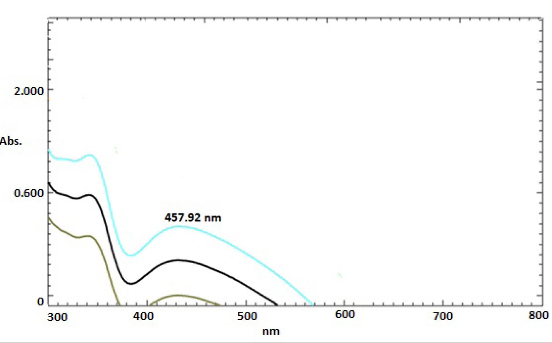
Figure 2.
UV-vis Spectra Showing the Maximum Wavelength Absorbance of Ma-AgNPs Synthesized through Mulberry Leaves. Note. UV-vis: Ultraviolet-visible; Ma-AgNPs: Synthesized silver nanoparticles
.
UV-vis Spectra Showing the Maximum Wavelength Absorbance of Ma-AgNPs Synthesized through Mulberry Leaves. Note. UV-vis: Ultraviolet-visible; Ma-AgNPs: Synthesized silver nanoparticles
The determination of the morphological appearance and distribution of the synthesized AgNP was made with the images obtained from the TEM and AFM micrographs given in Figures 3 and 4. TEM pictures illustrated that Ma-AgNPs have a morphological appearance that was predominantly spherical, and a small number of them seemed triangular. In AFM findings, Ma-AgNPs showed a distribution below 100 nm.26-29
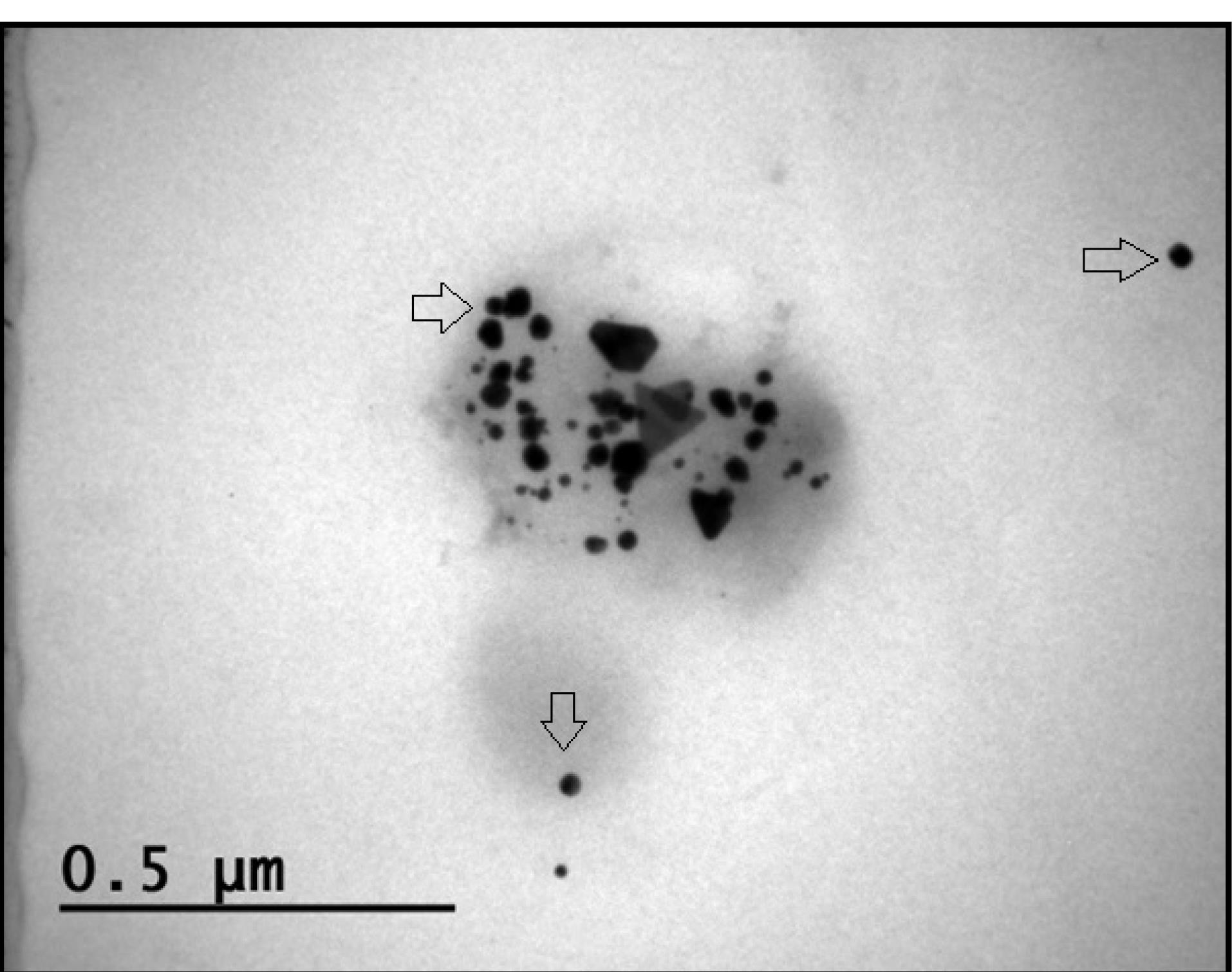
Figure 3.
TEM Picture Displaying the Ma-AgNPs’ Structural Appearances. Note. TEM: Transmissive electron microscope; Ma-AgNPs: Synthesized silver nanoparticles
.
TEM Picture Displaying the Ma-AgNPs’ Structural Appearances. Note. TEM: Transmissive electron microscope; Ma-AgNPs: Synthesized silver nanoparticles
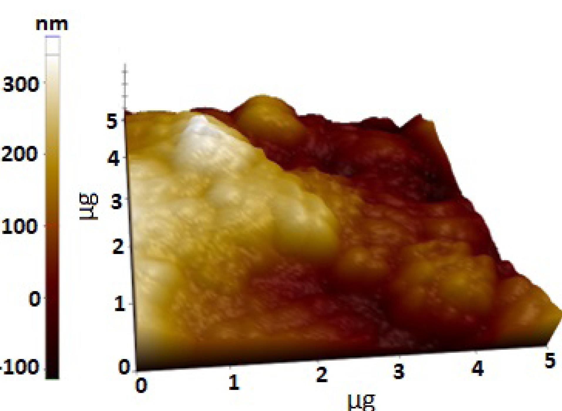
Figure 4.
AFM Micrograph Showing Topographic Distributions of Ma-AgNPs. Note. AFM: Atomic Force Microscopy; Ma-AgNPs: Synthesized silver nanoparticles
.
AFM Micrograph Showing Topographic Distributions of Ma-AgNPs. Note. AFM: Atomic Force Microscopy; Ma-AgNPs: Synthesized silver nanoparticles
The crystal structures and sizes of the particles synthesized using mulberry leaves were determined by the data received at 2θ via XRD. Bragg’s expansions at 111o, 200o, 220o, and 311o showed that Ma-AgNPs mostly have a central faceted cubic pattern (fcc),30-33 and the values obtained in the data were measured as 38.68, 44.54, 64.76, and 77.38, respectively (Figure 5). The crystal nano dimensions of Ma-AgNPs were calculated using the FWHM value of Debye-Sherer’s high peak, and the crystal size of Ma-AgNPs was 63.56 nm. In a green synthesis study in which different extraction processes were applied, it was calculated that the extracts have crystal sizes of 56 nm and 67 nm, respectively.34
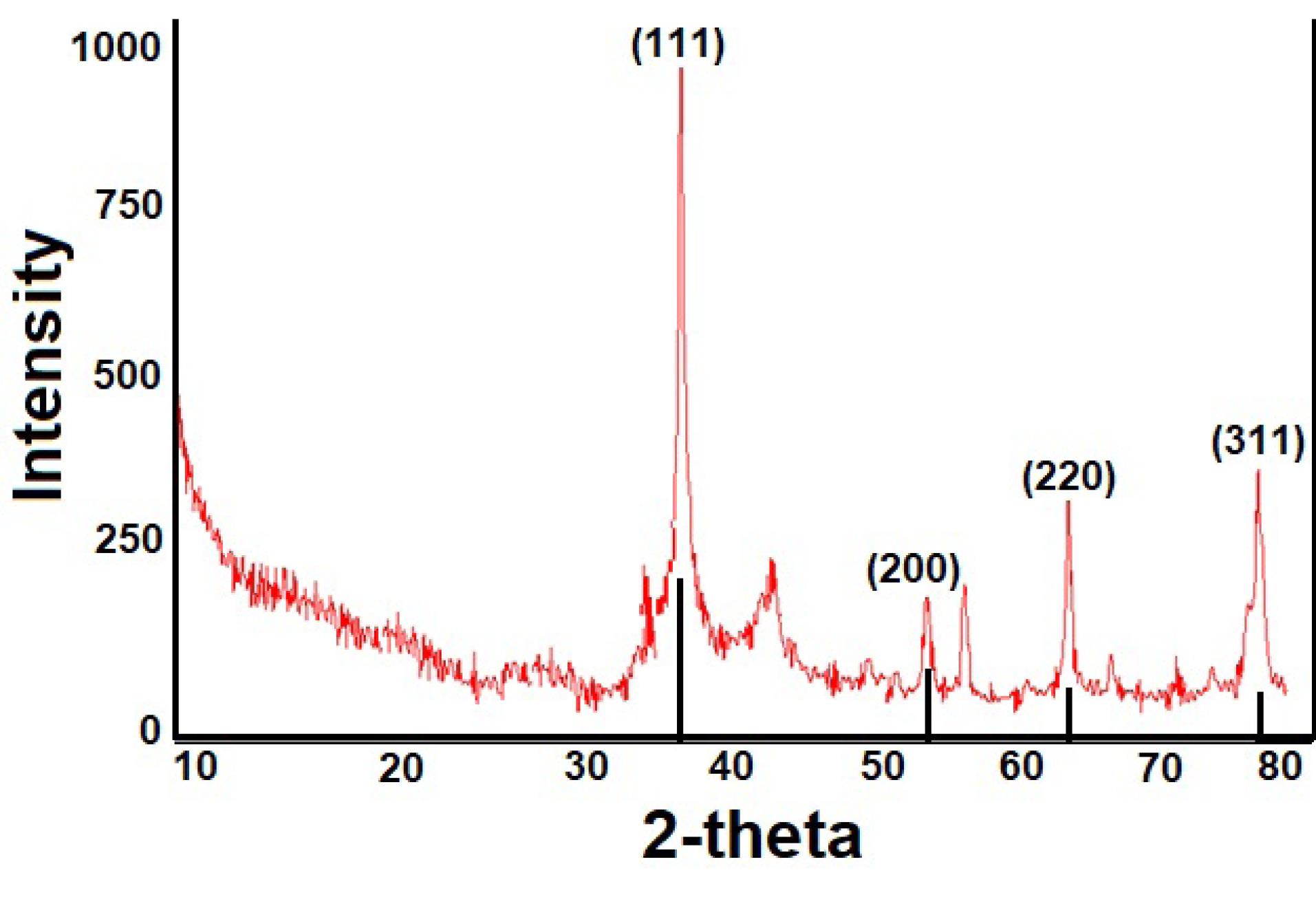
Figure 5.
XRD Data Showing the Crystal Structures of Ma-AgNPs Synthesized by the Leaf Extract. Note. XRD: X-ray diffraction; Ma-AgNPs: Synthesized silver nanoparticles
.
XRD Data Showing the Crystal Structures of Ma-AgNPs Synthesized by the Leaf Extract. Note. XRD: X-ray diffraction; Ma-AgNPs: Synthesized silver nanoparticles
Findings of Antibacterial Effect of Synthesized Silver Nanoparticles
Ma-AgNPs synthesized from mulberry leaves were determined on pathogenic Gram-negative and Gram-positive bacteria by MIC microdilution method. MIC values of Ma-AgNPs suppressing the growth of the strains are presented in Table 1. As can be seen, 0.25-0.50 µg mL-1 concentrations were found to have a suppressive effect on E. coli and S. aureus. These concentrations were lower than the commercial antibiotics used in practice (Table 1).
Table 1.
Concentrations of Ma-AgNPs, Antibiotics, and Silver Nitrate Solution Where They Exert an Antimicrobial Effect on the Growth of Microorganisms
|
Organism
|
AgNPs
µg mL-1 |
Silver Nitrate
µg mL-1 |
Antibiotic
µg mL-1 |
|
Staphylococcus aureus
|
0.50 |
4.00 |
2.00 |
|
Escherichia coli
|
0.25 |
2.00 |
2.00 |
Note. Ma-AgNPs: Synthesized silver nanoparticles.
Ma-AgNPs were ionized and exhibited considerable reactivity in liquid media. This reactivity was created by the engagement of electrostatic attraction force interactions with the microorganisms present in the same environment.35-39 By causing damage to microorganisms’ membrane structures, they enhanced the production of reactive oxygen species and altered the biomolecules’ structures, which caused a decline in their functions and the extinction of microorganisms.18,40 In this study, Ma-AgNPs were synthesized easily and quickly with the extract obtained by collecting the leaves of the M. alba L. plant in Orduzu, Malatya region. The outcomes of several microscopic and spectroscopic analyses were used to evaluate the characteristics of Ma-AgNPs. Ma-AgNPs can be produced as a powerful substitute, particularly in the struggle against microorganisms with multiple drug resistance.
Discussion
Rapid color change was a macroscopic finding indicating the formation of Ma-AgNPs.20-24 In addition to this finding given in Figure 1, the maximum absorbance bands at 457.92 nm taken with UV-vis were characteristic spectrums showing the presence of Ma-AgNPs with the formation of on the plasma surface rezonans (SPR).21-25 It was observed that Ma-AgNPs were mostly spherical in morphological appearance in TEM images, and they also showed triangular appearance, although a small number of them. In AFM findings, it was observed that they showed a distribution below 100 nm.28,29 Bragg’s expansions at 111o, 200o, 220o and 311o showed that Ma-AgNPs mostly have a central faceted cubic pattern (fcc).30-33 The values obtained in the data were measured as 38.68, 44.54, 64.76, and 77.38, respectively (Figure 4). The crystal nano dimensions of Ma-AgNPs were calculated using the FWHM value of Debye-Sherer’s high peak, and the crystal size of Ma-AgNPs was 63.56 nm. In a green synthesis study in which different extraction processes were applied, it was calculated that the extracts had crystal sizes of 56 nm and 67 nm, respectively.34 As seen in the findings, 0.25-0.50 µg mL-1 concentrations were found to have a suppressive effect on E. coli and S. aureus. These concentrations were lower than the commercial antibiotics used in practice (Table 1). Ma-AgNPs, In liquid medium are ionized and show high reactivity. They provide this reactivity by interacting with the microorganisms in the same environment with the electrostatic attraction force.35-39 They cause an increase in reactive oxygen species (ROS) by disrupting the membrane structure of microorganisms. They disrupt the structure and functions of RNA, DNA, and enzymes with high affinity for these species. The structures of biomolecules damaged by these species and as a result their functions deteriorate, resulting in the death of microorganisms.8,18,40
Conclusions
Green synthesis methods are a very popular area that aims to design safer and more sustainable processes without the use of toxic chemicals. The Ma-AgNPs had size distributions below 100 nm, an ultimate absorbance of 457.92 nm, and a mainly spherical shape. By defining the MIC, the antibacterial properties of pathogen gram-negative and positive bacteria have been identified by microdilution. Furthermore, Ma-AgNPs were discovered to be more successful than generic antibiotics, according to the data.
Acknowledgments
We would like to thank Batman University BAP unit for their support of this study. (BTÜBAP-2023-TBMYO-07).
Funding
None.
Data availability statement
Not applicable.
Ethical approval
Not applicable.
Consent for publication
Not applicable.
Conflict of interests
The authors declare that there is no conflict of interests.
References
- Baran MF, Saydut A, Umaz A. Silver nanomaterial synthesis and antimicrobial applications. Dicle Univ J Eng 2019; 10(2):689-95. [ Google Scholar]
- Baran MF, Saydut A. Gold nanomaterial synthesis and characterization. Dicle Univ J Eng 2019; 10(3):1033-40. [ Google Scholar]
- Baran MF. Synthesis and antimicrobial applications of silver nanoparticles from Artemisia absinthium plant. Biol Chem Res 2019; 6:96-103. [ Google Scholar]
- Ramkumar VS, Pugazhendhi A, Gopalakrishnan K, Sivagurunathan P, Saratale GD, Dung TNB. Biofabrication and characterization of silver nanoparticles using aqueous extract of seaweed Enteromorpha compressa and its biomedical properties. Biotechnol Rep (Amst) 2017; 14:1-7. doi: 10.1016/j.btre.2017.02.001 [Crossref] [ Google Scholar]
- Rauf A, Ahmad T, Khan A, Maryam Maryam, Uddin G, Ahmad B. Green synthesis and biomedicinal applications of silver and gold nanoparticles functionalized with methanolic extract of Mentha longifolia. Artif Cells Nanomed Biotechnol 2021; 49(1):194-203. doi: 10.1080/21691401.2021.1890099 [Crossref] [ Google Scholar]
- İpek P, Baran MF, Yildiz R, Hatipoğlu A. Biosynthesis of silver nanoparticles from Arum dioscoridis plant leaf aqueous extract: anticancer and antimicrobial properties. Int J Agric Environ Food Sci 2023; 7(2):399-407. doi: 10.31015/jaefs.2023.2.18 [Crossref] [ Google Scholar]
- Baran MF, Keskin C, Baran A, Kurt K, İpek P, Eftekhari A, et al. Green synthesis and characterization of selenium nanoparticles (Se NPs) from the skin (testa) of Pistacia vera L. (Siirt pistachio) and investigation of antimicrobial and anticancer potentials. Biomass Convers Biorefin. 2023. 10.1007/s13399-023-04366-8.
- Huq MA, Ashrafudoulla M, Rahman MM, Balusamy SR, Akter S. Green synthesis and potential antibacterial applications of bioactive silver nanoparticles: a review. Polymers (Basel) 2022; 14(4):742. doi: 10.3390/polym14040742 [Crossref] [ Google Scholar]
- Palanisamy S, Rajasekar P, Vijayaprasath G, Ravi G, Manikandan R, Marimuthu Prabhu N. A green route to synthesis silver nanoparticles using Sargassum polycystum and its antioxidant and cytotoxic effects: an in vitro analysis. Mater Lett 2017; 189:196-200. doi: 10.1016/j.matlet.2016.12.005 [Crossref] [ Google Scholar]
- Khan AU, Yuan Q, Khan ZUH, Ahmad A, Khan FU, Tahir K. An eco-benign synthesis of AgNPs using aqueous extract of longan fruit peel: antiproliferative response against human breast cancer cell line MCF-7, antioxidant and photocatalytic deprivation of methylene blue. J Photochem Photobiol B 2018; 183:367-73. doi: 10.1016/j.jphotobiol.2018.05.007 [Crossref] [ Google Scholar]
- Rani P, Kumar V, Singh PP, Matharu AS, Zhang W, Kim KH. Highly stable AgNPs prepared via a novel green approach for catalytic and photocatalytic removal of biological and non-biological pollutants. Environ Int 2020; 143:105924. doi: 10.1016/j.envint.2020.105924 [Crossref] [ Google Scholar]
- Ghatage MM, Mane PA, Gambhir RP, Parkhe VS, Kamble PA, Lokhande CD. Green synthesis of silver nanoparticles via Aloe barbadensis miller leaves: anticancer, antioxidative, antimicrobial and photocatalytic properties. Appl Surf Sci Adv 2023; 16:100426. doi: 10.1016/j.apsadv.2023.100426 [Crossref] [ Google Scholar]
- Gryn-Rynko A, Bazylak G, Olszewska-Slonina D. New potential phytotherapeutics obtained from white mulberry (Morus alba L) leaves. Biomed Pharmacother 2016; 84:628-36. doi: 10.1016/j.biopha.2016.09.081 [Crossref] [ Google Scholar]
- Eren A, Baran MF. Green synthesis, characterization and antimicrobial activity of silver nanoparticles (AgNPs) from maize (Zea mays L). Appl Ecol Environ Res 2019; 17(2):4097-105. doi: 10.15666/aeer/1702_40974105 [Crossref] [ Google Scholar]
- Atalar MN, Baran A, Baran MF, Keskin C, Aktepe N, Yavuz Ö. Economic fast synthesis of olive leaf extract and silver nanoparticles and biomedical applications. Part Sci Technol 2022; 40(5):589-97. doi: 10.1080/02726351.2021.1977443 [Crossref] [ Google Scholar]
- Aktepe N, Bütüner H, Baran A, Baran MF, Keskin C. Synthesis, characterization and evaluation of antimicrobial activities of silver nanoparticles obtained from Rumex acetosella L(Sorrel) plant. Int J Agric Environ Food Sci 2022; 6(4):522-9. doi: 10.31015/jaefs.2022.4.4 [Crossref] [ Google Scholar]
- Patil MP, Seong YA, Kim JO, Seo YB, Kim GD. Synthesis of silver nanoparticles using aqueous extract of Cuscuta japonica seeds and their antibacterial and antioxidant activities. Inorg Chem Commun 2021; 134:109035. doi: 10.1016/j.inoche.2021.109035 [Crossref] [ Google Scholar]
- Emmanuel R, Palanisamy S, Chen SM, Chelladurai K, Padmavathy S, Saravanan M. Antimicrobial efficacy of green synthesized drug blended silver nanoparticles against dental caries and periodontal disease causing microorganisms. Mater Sci Eng C 2015; 56:374-9. doi: 10.1016/j.msec.2015.06.033 [Crossref] [ Google Scholar]
- Raghavendra VB, Shankar S, Govindappa M, Pugazhendhi A, Sharma M, Nayaka SC. Green synthesis of zinc oxide nanoparticles (ZnO NPs) for effective degradation of dye, polyethylene and antibacterial performance in waste water treatment. J Inorg Organomet Polym Mater 2022; 32(2):614-30. doi: 10.1007/s10904-021-02142-7 [Crossref] [ Google Scholar]
- Sattari R, Khayati GR, Hoshyar R. Biosynthesis of silver–silver chloride nanoparticles using fruit extract of Levisticum officinale: characterization and anticancer activity against MDA-MB-468 cell lines. J Clust Sci 2021; 32(3):593-9. doi: 10.1007/s10876-020-01818-3 [Crossref] [ Google Scholar]
- Jebril S, Khanfir Ben Jenana R, Dridi C. Green synthesis of silver nanoparticles using Melia azedarach leaf extract and their antifungal activities: in vitro and in vivo. Mater Chem Phys 2020; 248:122898. doi: 10.1016/j.matchemphys.2020.122898 [Crossref] [ Google Scholar]
- Acay H, Baran MF. Biosynthesis and characterization of silver nanoparticles using king oyster (Pleurotus eryngii) extract: effect on some microorganisms. Appl Ecol Environ Res 2019; 17(4):9205-14. [ Google Scholar]
- Mani M, Harikrishnan R, Purushothaman P, Pavithra S, Rajkumar P, Kumaresan S. Systematic green synthesis of silver oxide nanoparticles for antimicrobial activity. Environ Res 2021; 202:111627. doi: 10.1016/j.envres.2021.111627 [Crossref] [ Google Scholar]
- Mamdooh NW, Naeem GA. Green synthesis, characterization and biological activity of silver nanoparticles using Ruta leaf extract. J Phys Conf Ser 2021; 1999(1):012050. doi: 10.1088/1742-6596/1999/1/012050 [Crossref] [ Google Scholar]
- Arumai Selvan D, Mahendiran D, Senthil Kumar R, Kalilur Rahiman A. Garlic, green tea and turmeric extracts-mediated green synthesis of silver nanoparticles: phytochemical, antioxidant and in vitro cytotoxicity studies. J Photochem Photobiol B 2018; 180:243-52. doi: 10.1016/j.jphotobiol.2018.02.014 [Crossref] [ Google Scholar]
- Swamy MK, Akhtar MS, Mohanty SK, Sinniah UR. Synthesis and characterization of silver nanoparticles using fruit extract of Momordica cymbalaria and assessment of their in vitro antimicrobial, antioxidant and cytotoxicity activities. Spectrochim Acta A Mol Biomol Spectrosc 2015; 151:939-44. doi: 10.1016/j.saa.2015.07.009 [Crossref] [ Google Scholar]
- Kumar V, Gundampati RK, Singh DK, Bano D, Jagannadham MV, Hasan SH. Photoinduced green synthesis of silver nanoparticles with highly effective antibacterial and hydrogen peroxide sensing properties. J Photochem Photobiol B 2016; 162:374-85. doi: 10.1016/j.jphotobiol.2016.06.037 [Crossref] [ Google Scholar]
- Shaheen TI, Abd El Aty AA. In-situ green myco-synthesis of silver nanoparticles onto cotton fabrics for broad spectrum antimicrobial activity. Int J Biol Macromol 2018; 118(Pt B):2121-30. doi: 10.1016/j.ijbiomac.2018.07.062 [Crossref] [ Google Scholar]
- Pinzaru I, Coricovac D, Dehelean C, Moacă EA, Mioc M, Baderca F. Stable PEG-coated silver nanoparticles – a comprehensive toxicological profile. Food Chem Toxicol 2018; 111:546-56. doi: 10.1016/j.fct.2017.11.051 [Crossref] [ Google Scholar]
- Acay H, Baran MF, Eren A. Investigating antimicrobial activity of silver nanoparticles produced through green synthesis using leaf extract of common grape (Vitis Vinifera). Applied Ecology and Environmental Research 2019; 17(2):4539-46. doi: 10.15666/aeer/1702_45394546 [Crossref] [ Google Scholar]
- Wongpreecha J, Polpanich D, Suteewong T, Kaewsaneha C, Tangboriboonrat P. One-pot, large-scale green synthesis of silver nanoparticles-chitosan with enhanced antibacterial activity and low cytotoxicity. Carbohydr Polym 2018; 199:641-8. doi: 10.1016/j.carbpol.2018.07.039 [Crossref] [ Google Scholar]
- Anandalakshmi K. Green synthesis, characterization and antibacterial activity of silver nanoparticles using Chenopodium album leaf extract. Indian J Pure Appl Phys 2021; 59(6):456-61. doi: 10.56042/ijpap.v59i6.32055 [Crossref] [ Google Scholar]
- Singh A, Sharma B, Deswal R. Green silver nanoparticles from novel Brassicaceae cultivars with enhanced antimicrobial potential than earlier reported Brassicaceae members. J Trace Elem Med Biol 2018; 47:1-11. doi: 10.1016/j.jtemb.2018.01.001 [Crossref] [ Google Scholar]
- Ali BM. Eco-friendly synthesis of silver nanoparticles from crust of Cucurbita maxima L(red pumpkin). EurAsian J BioSci 2020; 14(2):2829-33. [ Google Scholar]
- Chung IM, Park I, Seung-Hyun K, Thiruvengadam M, Rajakumar G. Plant-mediated synthesis of silver nanoparticles: their characteristic properties and therapeutic applications. Nanoscale Res Lett 2016; 11(1):40. doi: 10.1186/s11671-016-1257-4 [Crossref] [ Google Scholar]
- Zhang Y, Yang D, Kong Y, Wang X, Pandoli O, Gao G. Synergetic antibacterial effects of silver nanoparticles@Aloe vera prepared via a green method. Nano Biomed Eng 2010; 2(4):252-7. doi: 10.5101/nbe.v2i4.p252-257 [Crossref] [ Google Scholar]
- Sinha SN, Paul D, Halder N, Sengupta D, Patra SK. Green synthesis of silver nanoparticles using fresh water green alga Pithophora oedogonia (Mont) Wittrock and evaluation of their antibacterial activity. Appl Nanosci 2015; 5(6):703-9. doi: 10.1007/s13204-014-0366-6 [Crossref] [ Google Scholar]
- Aina AD, Owolo O, Adeoye-Isijola M, Aina FO, Favour O, Adewumi AG. Almond leaves for the one-pot biofabrication of silver nanoparticles: characterization and larvicidal application. Int J Sci Res Publ 2018; 8(11):703-11. doi: 10.29322/IJSRP.8.11.2018.p8378 [Crossref] [ Google Scholar]
- Ahmed KB, Raman T, Veerappan A. Future prospects of antibacterial metal nanoparticles as enzyme inhibitor. Mater Sci Eng C 2016; 68:939-47. doi: 10.1016/j.msec.2016.06.034 [Crossref] [ Google Scholar]
- Shao Y, Wu C, Wu T, Yuan C, Chen S, Ding T. Green synthesis of sodium alginate-silver nanoparticles and their antibacterial activity. Int J Biol Macromol 2018; 111:1281-92. doi: 10.1016/j.ijbiomac.2018.01.012 [Crossref] [ Google Scholar]